
Male Breast Cancer
Staging
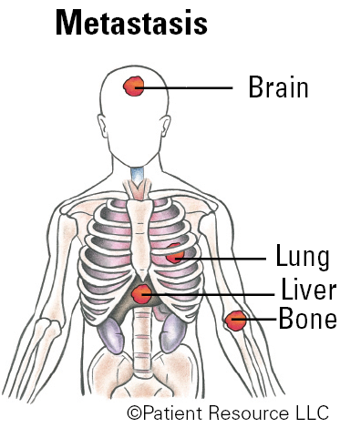
The results of a biopsy, imaging scans and genomic testing are used to classify and stage breast cancer according to the tumor, node and metastasis (TNM) system developed by the American Joint Committee on Cancer (AJCC). The system includes the tumor (T) size, cancer cells found in nearby lymph nodes (N), and cancer that has metastasized (M), or spread, to other parts of the body, such as the bones, brain, liver or lungs (see Table 1).
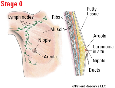
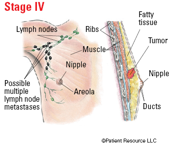
After breast cancer is classified, it is staged (see Table 2 and Figure 1). Stage 0 refers to ductal carcinoma in situ (DCIS) breast cancer, and Stage IV represents breast cancer that has spread beyond the breast and lymph nodes into distant organs or bone.
Before a final stage is determined, many factors are considered: tumor grade; biomarkers, including the tumor’s estrogen receptor (ER), progesterone receptor (PR) and human epidermal growth factor receptor-2 (HER2) status; and molecular and genetic changes in cancer tissue identified in multigene panels such as MammaPrint, Oncotype DX, PAM 50 (Prosigna) and the Breast Cancer Index.
The hormone-related biomarkers ER and PR send signals to special receptor proteins inside normal breast cells and some breast cancer cells (those that carry the ER and/or PR biomarkers) to “turn on” the growth of cells. As a result, breast cancers are classified according to the presence (ER+/PR+) or absence (ER-/PR-) of these hormone receptors in the cells, and the amount (or expression) of receptors. Most breast cancers in men are hormone receptor-positive, which means the growth of cancer cells is stimulated by estrogen and/or progesterone, both of which are found in men.
Approximately 20 percent of all breast cancers make extra copies of HER2, which encodes a growth-promoting protein. Breast cancers with too much of this protein tend to grow and spread more aggressively. Breast cancer that does not express either of the hormone receptors or the HER2 receptor is referred to as triple-negative breast cancer (TNBC), an aggressive form of breast cancer that is rarely diagnosed in men.
Determining whether you have hereditary breast cancer is important for your family members. The BReast CAncer 1 (BRCA1) and BReast CAncer 2 (BRCA2) genes are the most common hereditary susceptibility genes, and your doctor may test for others. Family members that have inherited abnormalities in the BRCA1 or BRCA2 genes have an increased likelihood of developing breast cancer and/or ovarian cancer.
Newly-diagnosed breast cancer patients found to have a BRCA mutation face an increased risk of another new breast cancer. As a result, the presence of inherited mutations in the BRCA1 and BRCA2 genes or other cancer-susceptibility genes may influence decisions regarding cancer prevention (prophylactic) surgery (removal of the breasts). The discovery of these mutations may also lead to different systemic treatments.
Keep in mind that having an inherited mutation does not mean you will automatically or definitely develop cancer; it means the risk is increased and you can explore ways to lower it, such as preventive surgery, medication or lifestyle changes. Frequent screenings will most likely result in early detection.
Risk factors that suggest a person carries the BRCA mutation include:
- Family history of any cancer, especially rare, including male breast cancer
- Cancer at an early age
- Multiple cancers in one relative
- Certain ancestry, such as Ashkenazi Jewish heritage
- Triple-negative breast cancer
Table 1. TNM System for Classifying Breast Cancers
| Category | Definition |
| Tumor (T) | |
| TX | Primary tumor cannot be assessed. |
| T0 | No evidence of primary tumor. |
| Tis (DCIS) | Ductal carcinoma in situ. |
| Tis (Paget) | Paget disease of the nipple NOT associated with invasive carcinoma and/or carcinoma in situ (DCIS) in the underlying breast parenchyma (tissue). |
| T1 | Tumor ≤ (not more than) 20 mm in greatest dimension. |
| T1mi | Tumor ≤ (not more than) 1 mm in greatest dimension. |
| T1a | Tumor > (more than) 1 mm but ≤ (not more than) 5 mm in greatest dimension. |
| T1b | Tumor > (more than) 5 mm but ≤ (not more than) 10 mm in greatest dimension. |
| T1c | Tumor > (more than) 10 mm but ≤ (not more than) 20 mm in greatest dimension. |
| T2 | Tumor > (more than) 20 mm but ≤ (not more than) 50 mm in greatest dimension. |
| T3 | Tumor > (more than) 50 mm in greatest dimension. |
| T4 | Tumor of any size with direct extension to the chest wall and/or to the skin (ulceration or macroscopic nodules). |
| T4a | Extension to the chest wall. |
| T4b | Ulceration and/or ipsilateral (on the same side) macroscopic satellite nodules and/or edema (including peau d’orange) of the skin that does not meet the criteria for inflammatory carcinoma. |
| T4c | Both T4a and T4b are present. |
| T4d | Inflammatory carcinoma. |
| Node (N) | |
| pNX | Regional lymph nodes cannot be assessed. |
| pN0 | No regional lymph node metastasis identified or ITCs (isolated tumor cells) only. |
| pN0(i+) | ITCs (isolated tumor cells) only (malignant cell clusters no larger than 0.2 mm) in regional lymph node(s). |
| pN0(mol+) | Positive molecular findings by reverse transcriptase polymerase chain reaction (RT-PCR); no ITCs (isolated tumor cells) detected. |
| pN1 | Micrometastases; or metastases in 1-3 axillary (armpit) lymph nodes; and/or clinically negative internal mammary nodes with micrometastases or macrometastases by sentinel lymph node biopsy. |
| pN1mi | Micrometastases (approximately 200 cells, larger than 0.2 mm, but none larger than 2.0 mm). |
| pN1a | Metastases in 1-3 axillary (armpit) lymph nodes, at least one metastasis larger than 2.0 mm. |
| pN1b | Metastases in ipsilateral (on the same side) internal mammary sentinel nodes, excluding ITCs (isolated tumor cells). |
| pN1c | pN1a and pN1b combined. |
| pN2 | Metastases in 4-9 axillary (armpit) lymph nodes; or positive ipsilateral (on the same side) internal mammary lymph nodes by imaging in the absence of axillary lymph node metastases. |
| pN2a | Metastases in 4-9 axillary (armpit) lymph nodes (at least one tumor deposit larger than 2.0 mm). |
| pN2b | Metastases in clinically detected internal mammary lymph nodes with or without microscopic confirmation; with pathologically negative axillary (armpit) nodes. |
| pN3 |
Metastases in 10 or more axillary (armpit) lymph nodes;
or in infraclavicular (below the clavicle) (Level III axillary) lymph nodes; or positive ipsilateral (on the same side) internal mammary lymph nodes by imaging in the presence of one or more positive Level I, II axillary lymph nodes; or in more than three axillary lymph nodes and micrometastases or macrometastases by sentinel lymph node biopsy in clinically negative ipsilateral internal mammary lymph nodes; or in ipsilateral supraclavicular (above the clavicle) lymph nodes. |
| pN3a |
Metastases in 10 or more axillary (armpit) lymph nodes (at least one tumor deposit larger than 2.0 mm);
or metastases to the infraclavicular (below the clavicle) (Level III axillary) lymph nodes. |
| pN3b | pN1a or pN2a in the presence of cN2b (positive internal mammary nodes by imaging);
or pN2a in the presence of pN1b. |
| pN3c | Metastases in ipsilateral (on the same side) supraclavicular (above the clavicle) lymph nodes. |
| Note: (sn) and (f) suffixes should be added to the N category to denote confirmation of metastasis by sentinel node biopsy or FNA/core needle biopsy respectively, with NO further resection of nodes. | |
| Metastasis (M) | |
| M0 | No clinical or radiographic evidence of distant metastases. |
| cM0(i+) | No clinical or radiographic evidence of distant metastases in the presence of tumor cells or deposits no larger than 0.2 mm detected microscopically or by molecular techniques in circulating blood, bone marrow, or other nonregional nodal tissue in a patient without symptoms or signs of metastases. |
| cM1 | Distant metastases detected by clinical and radiographic means. |
| pM1 | Any histologically proven metastases in distant organs; or if in non-regional nodes, metastases greater than 0.2 mm. |
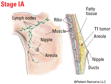
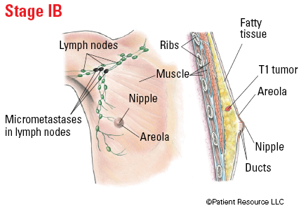
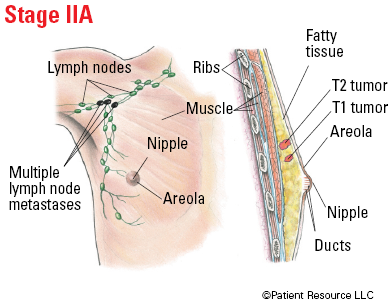
Table 2. Stages of Breast Cancer
| Stage | TNM Classification |
| 0 | Tis, N0, M0 |
| IA | T1, N0, M0 |
| IB | T0 or T1, N1mi, M0 |
| IIA |
T0 or T1, N1, M0
T2, N0, M0 |
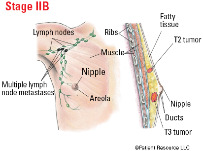
| IIB |
T2, N1, M0
T3, N0, M0 |
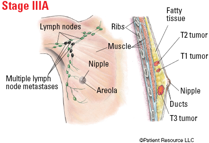
| IIIA |
T0-T3, N2, M0
T3, N1, M0 |
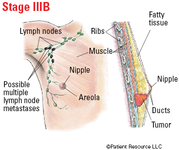
| IIIB | T4, N0-N2, M0 |
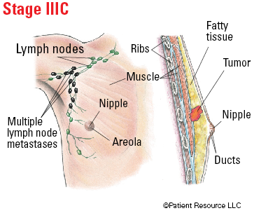
| IIIC | Any T, N3, M0 |
| IV | Any T, Any N, M1 |
Illustrated Stages of Breast Cancer (Male)