

Gynecologic Cancer
Imaging Techniques
 Imaging studies can play an important role in diagnosing gynecologic cancers. They can help define the size and location of the tumor as well as the extent of disease, which are all essential pieces of information in determining the stage of the cancer. Imaging studies use minimal radiation and typically cause no pain or side effects, but some tests may cause minor discomfort (Table 1).
X-rays
Standard X-rays are often the first imaging study done because of their convenience and low cost. X-rays are primarily used to rule out the spread of cancer (metastasis) to the lungs or bones, which are common places for cancer to spread (metastatic sites). If an X-ray shows evidence of metastasis, another imaging study is usually needed to confirm the findings.
Ultrasound
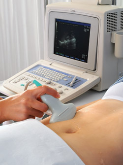
Ultrasound of the pelvis and/or abdomen can help better define a tumor in the reproductive system or abdomen. Transvaginal ultrasound is often done to evaluate suspected gynecologic cancers.
Bone scan
A bone scan is typically done to determine whether the cancer has spread to the bone. An advantage of this test is that it provides images of all the bones in the body at the same time.
Computerized tomography (CT)
CT scans are usually done to confirm the findings on a chest X-ray or to evaluate the abdominal organs or the brain for signs of metastasis. A CT scan provides three-dimensional, cross-sectional X-ray images, showing more details than a standard X-ray.
Magnetic resonance imaging (MRI)
MRI of the pelvis is particularly helpful for examining tumors in that area of the body. MRI may also be done to look for metastasis in other parts of the body and is especially useful for detecting brain metastasis.
Positron emission tomography (PET)
PET scans are rarely used in evaluating early-stage gynecologic cancers, but they may be helpful for assessing advanced disease. In addition, a combination of PET and CT is increasingly used to assess response to chemotherapy and/or hormone therapy as well.
Table 1. Imaging studies: What to expect
| Imaging study | How it's done |
| Standard X-rays | Chest X-rays are usually taken while you’re standing with your chest against a plate, and abdominal X-rays are usually taken while you’re lying on a table. You may be asked to change positions if more than one view is needed, and you must keep still and hold your breath for a few seconds when the X-ray picture is taken. |
| Ultrasound | A small, microphone-like instrument (transducer) is moved over the part of your body being studied. It sends out sound waves, which bounce off your body tissues and create echoes. The echoes are then converted into images that provide an outline of that area of the body. With transvaginal ultrasound, the transducer is inserted into the vagina to provide images of pelvic organs, such as the uterus and ovaries. |
| Bone scan | A small amount of a low-level radioactive substance is swallowed, inhaled or injected into a vein in your arm. This substance collects in areas of metastatic disease in bone. You will then lie on a table (for about 30 minutes), and a special camera will record images of areas with an increased amount of the radioactive substance. Most of the substance will pass through your body within one day and be completely gone within two days. This radioactivity poses no risk to you or others around you. |
| Computerized tomography (CT) | The CT scanner takes several pictures of your organs as the table on which you’re lying moves slowly through the scanner. You will need to lie very still during this process. A contrast material (dye) may be injected into a vein in your arm (or given by mouth) before the test to enhance the quality of the images. This dye may cause a brief sense of warmth or flushing in your body; this feeling is normal. |
| Magnetic resonance imaging (MRI) | Images are produced through radio waves and a powerful magnet linked to a computer. Although MRI is painless, you will need to lie still on a table within the tube of the MRI machine, which makes loud, repetitive clicking noises. Tell your doctor if you are uncomfortable in enclosed spaces so that steps can be taken to manage your anxiety. Contrast material may be used to enhance the images. |
| Positron emission tomography (PET) | A small amount of a radioactive sugar (or other radioactive substance) is injected into a vein in your arm. Cancer cells use a high amount of energy, so they will absorb large quantities of the radioactive sugar. A special camera will detect this absorption and provide an image of the tumor. |
